Leaders across Research, Translational Medicine and the Clinic rely on RareCyte as a one-stop resource for Spatial Biology and Liquid Biopsy solutions.
Our highly multiplexed fluorescent imaging systems, reagents and contract services empower investigators to unlock spatial context for key biomarkers and develop therapeutics and companion diagnostics though rare cell analysis.
 |
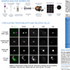
Webinar: Unlocking Tissue Cytometry with Orion™ |
 |
Pioneering circulating trophoblast research featured at the 2024 SMFM Pregnancy Meeting |
 |
Nature Oncogene publication: Neoadjuvant botensilimab plus balstilimab response pattern investigated |
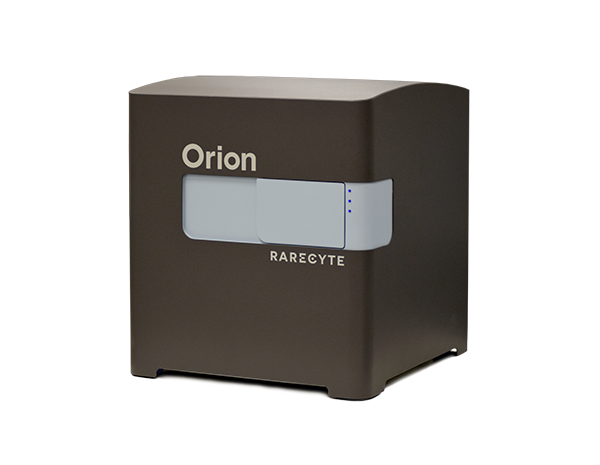
Break barriers with the fastest path to whole-slide, highly multiplexed biomarker imaging data. Orion brings biomarker depth and flexibility with the convenience of a single-step stain and image process.
Investigate circulating tumor cell (CTC) biomarker expression and mutational status using a non-invasive liquid biopsy. Obtain sensitive, accurate, and reproducible results utilizing a complete end-to-end workflow, from blood collection to single-cell isolation.
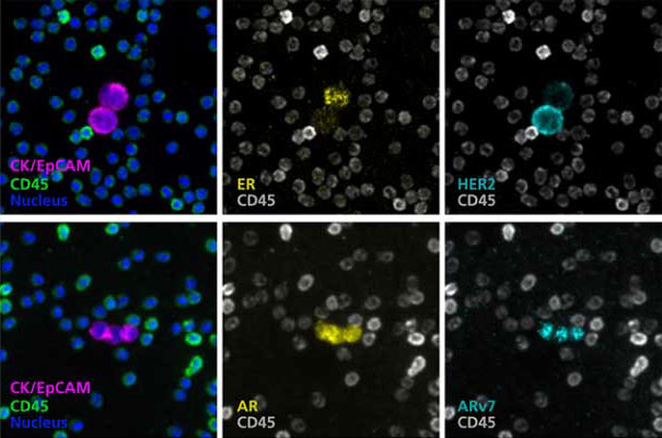
RareCyte enables class-leading performance in CTC applications and Spatial Biology solutions supporting therapy selection, disease-monitoring, and global research and clinical trials.
"Using their liquid biopsy imaging technology, RareCyte was able to develop a custom assay. Throughout the process, the RareCyte team was collaborative and insightful, showing true dedication to the project, resulting in the development of a fully validated assay."
Erica Tobin
Associate Director, Oncology
KSQ Therapeutics, Inc.
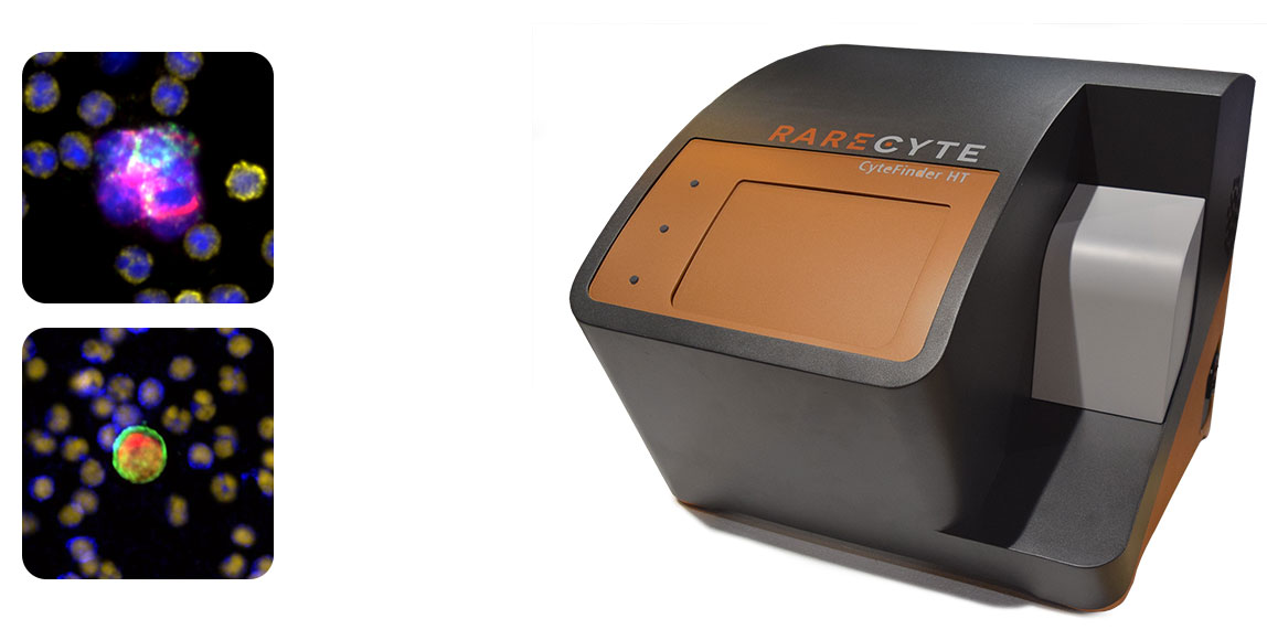
Enable rapid, high-throughput fluorescence imaging with CyteFinder II HT, and rare cell identification with single-cell retrieval with the CyteFinder II.
 |
Webinar: Use of open-source software for quantitative analysis of multiplex images |
 |
SOPHiA GENETICS Enhances RareCyte Precision Biology Services Portfolio |
 |
Measuring versus reading - Yale CLIA test transforming the field of quantitative pathology |
Need a refill of AccuCyte BCTs or a new biomarker panel kit? Select from our list of products to begin the order process.
For instrument orders and service programs, please Contact Us directly.