Webinars
Register for these upcoming webinars or view past webinars on demand to learn more about RareCyte, and how our products and services are making an impact today!
On Demand Webinars
Spatial Biomarker Panels for Clinical Trials
Most immuno-oncology drugs work by altering the tumor microenvironment to recruit and activate immune cells. To understand how these drugs work and how patients respond, researchers need the spatial context of several biomarkers—typically 8 to 18—to see how cells interact within the tumor. Traditional multiplex immunofluorescence (mIF) methods can’t capture this complexity at scale.
Through a case study and technology walk-through, this webinar highlights the barriers that have been broken in mIF that now enable flexible development of high performance high-plex biomarker panels, and application of those panels at the scale necessary for deriving valuable quantitative spatial insights from large cohort clinical trials.
Speaker
Jennifer Bordeaux, PhD
Associate Director, Digital Pathology Services
Navigate Biopharma Services, Inc.
Jennifer Bordeaux, Ph.D. is a strategic leader with over a decade of industry experience in digital pathology. As Associate Director of Digital Pathology Solutions at Navigate Biopharma Services, she leads a team of scientists driving the adoption of multiplexed fluorescence immunochemistry assays to support clinical trial programs. Her expertise spans assay development, image analysis, and biomarker validation, with a strong focus on operational efficiency and scientific innovation. Jennifer earned her Ph.D. in Experimental Pathology from Yale University, where her research centered on biomarker validation in breast cancer using AQUA technology. She is a published author and frequent presenter in the field of quantitative pathology.

Speaker
Tad George, PhD
Senior VP, Biology R&D
RareCyte Inc.
Tad has over 15 years of startup experience dedicated to creating scientific markets for novel instrumentation platforms that span basic research, drug discovery and clinical applications. Prior to joining RareCyte, Tad has held similar positions at Biodesy, Inc. and DVS Sciences, and was Director of Biology at Amnis Corporation. Tad completed his B.A. in Biochemistry from the Univ. of Texas at Austin, Ph.D. in Immunology from UT Southwestern Medical Center at Dallas, and post-doctoral training at Immunex Corp. in Seattle.
Bridging the Discovery-to-Translational Gap in Spatial Biology
Speakers:
Simon Goldstein
Microscopy Specialist II, La Jolla Institute for Immunology
Tad George, PhD
Senior VP, Biology R&D, RareCyte Inc.
Achieving reliable spatial biology results begins with confident antibody validation, robust multiplex panel design, and high-quality tissue preparation. In this webinar, Simon Goldstein from La Jolla Institute for Immunology, will share strategies to streamline multiplex immunofluorescence (IF) workflows and improve data consistency. You’ll learn practical tips for validating antibody performance and building custom panels that perform seamlessly across diverse tissue types and research goals.
Bridging the Discovery-to-Translational Gap in Spatial Biology
Speakers:
Zhihong Chen, PhD
CTO, OCCAM Immune, at Icahn School of Medicine, Mount Sinai Hospital, New York
Tad George, PhD
Sr VP Bio R&D, RareCyte
Spatial biology is uncovering new biomarker candidates, yet a major challenge remains: how to move from broad high-plex discovery to clinically relevant panels suitable for translational and clinical research workflows. Watch this live session for direct Q&A with two spatial biology experts and see how capturing more channels per imaging round enables you to perform high-plex discovery, then streamline to single-round panels optimized for large cohort studies.

Exploring new frontiers in pancreatic cancer treatment with spatial biology
Speakers:
Yana Zavros, PhD
Professor, University of Georgia School of Medicine
Tad George, PhD
Sr VP Bio R&D, RareCyte
Pancreatic ductal adenocarcinoma (PDAC) remains one of the deadliest cancers, with limited treatment options and a high recurrence rate. Emerging research suggests that the tumor microenvironment (TME) and metastatic site play a critical role in therapy response. In this webinar, Yana Zavros will discuss how she combined spatial biology and patient-derived organoids (PDOs) to uncover a distinct cell population driving therapy resistance and disease recurrence in PDAC.

Breaking Barriers in Spatial Proteomics with Single-Round Imaging
Speaker
Tad George, PhD
Sr VP Bio R&D, RareCyte
A challenge in multiplex immunofluorescence imaging is the trade-off between multiplexing and throughput. The Orion™ platform overcomes this limitation with 20-channel single-round spatial protein profiling, enabling researchers to perform high-plex spatial biomarker analysis at speeds suitable for large-scale studies.
In this webinar, you'll get an in-depth look at Orion’s capabilities, a walkthrough of its single-round workflow, and customer case studies showcasing key applications. Discover how the Orion platform is setting a new standard in spatial proteomics.

Accelerating Data and Throughput in Spatial Imaging: A Core Facility Perspective.
Speaker
Zbigniew Mikulski, PhD
Director of Microscopy and Instructor, La Jolla Institute for Immunology
In this webinar, Dr. Mikulski will share best practices from working on over 18 different projects, and how the Orion platform for spatial biology facilitated a fast, low run cost solution that works with FFPE samples. Dr. Mikulski will also discuss practices that other labs might adopt to further improve workflow efficiency and cost-effectiveness.

New discoveries in HER2 detection: A novel method of multimodal detection of HER2 from a single tube of blood
Speaker
Eszter Papp, PhD
R&D Product Manager, CellCarta
In this webinar, Dr. Papp presents a novel method for oncosome detection developed by the CellCarta team using the RareCyte CTC platform. She also presents findings from a proof-of-concept study that combined data from oncosomes, CTCs, and cfDNA analysis to identify metastatic breast cancer patients with elevated HER2 expression.

Use of open-source software for quantitative analysis of multiplex images
Speaker
Dr. Sara McArdle
La Jolla Institute of Immunology
Dr. McArdle discusses her use of open-source software for quantitative spatial analysis of tissue images. She reviews how multiplex immunostaining is a key method for understanding spatial context of the tissue microenvironment and statistical quantification of cells.

Multimodal tissue imaging and machine learning to advance precision medicine
Speaker
Dr. Peter K Sorger
Harvard Medical School
The effective treatment of cancer and many other diseases is increasingly dependent on a precision approach in which the quantification of molecular features at the level of individual patients is used to guide treatment plans. However, cancer diagnosis and staging are currently performed primarily via direct examination of biopsy and resection specimens by histopathologists. These classical methods provide insufficient molecular insight to guide the use of targeted and immunotherapies even when supplemented by knowledge of tumor genotypes.

Spatial analysis and high-plex immunofluorescence to study human pancreata in type 1 diabetes
Speaker
Dr. Estefania Quesada-Masachs
Instructor, La Jolla Institute for Immunology
In this webinar, Dr. Quesada-Masachs will explain the image analysis pipeline that she has developed and applied to study whole tissue pancreatic samples of donors with type 1 diabetes (T1D). Those samples were previously stained and acquired with the high-plex Orion™ platform. New methods for spatial image analysis are providing a better understanding of local processes occurring in many human diseases, revolutionizing those scientific fields.

Spatial Biology: Applying Cellular Phenotyping in Health and Disease
Speaker
Mickael Meyrand, Field Application Scientist, RareCyte, Inc
Tissue consists of heterogeneous cell types, each with diverse functions and functional states, where spatial organization can impact patient health status. Understanding such spatial context at the subcellular level is often challenging due to low resolution imaging, throughput, and simultaneous target assessment. Orion™ technology can resolve these challenges, providing a fast path to whole slide, multiplexed, and high resolution biomarker imaging data

Circulating Tumor Cells Circa 2022
Speaker
Daniel E. Sabath, MD, PhD
University of Washington Department of Laboratory Medicine and Pathology
Detecting small numbers of tumor cells is technically challenging, especially for patients with solid tumors, where very small numbers of tumor cells are present in the peripheral blood, even in patients with widely metastatic disease. In this presentation, Dr. Sabath will describe various technologies for detecting circulating tumor cells and data suggesting clinical utility. In particular, his laboratory's clinical validation of a circulating tumor cell assay using density gradient centrifugation to enrich for circulating tumor cells. Dr. Sabath will also discuss possible future applications of this technology.
Unlock Spatial Biology with Orion Technology: Explore the possibilities for research and clinical applications
Speaker
Mickael Meyrand, Field Application Scientist, RareCyte, Inc
Tissue consists of heterogenous cell types, each with diverse functions and functional states, arranged spatially in a way that impacts patient health status. Resolving this complexity at the subcellular level has historically been challenged by image resolution, the number of targets that can be simultaneously assessed, and throughput. Orion™ technology breaks these barriers by providing the fastest path to whole slide, high-plex imaging which will be shown in this webinar.
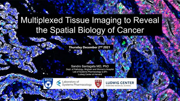
Multiplexed Tissue Imaging using the Orion Platform to Reveal the Spatial Biology of Cancer
Speaker
Dr. Sandro Santagata, MD PhD
Associate Professor Pathology, Department of Pathology, at Brigham and Women's Hospital
Recent developments in tissue imaging technologies have made available new tools that promise to advance our understanding of human disease and help guide the development and implementation of new therapies. This interactive webinar will focus on the use of multiplexed antibody-based tissue imaging of multiple antigens at single cell resolution using both cyclic immunofluorescence (CyCIF) and Orion high-plex imaging and the use of this data to identify cell lineages and states (molecular phenotypes) and map their spatial organization and interactions at different scales.

Patient Treatment Selection & Disease Monitoring by CTC enumeration
Speakers
Dr. Qingyan Au, Director of Multiplexing, NeoGenomics
Dr. Pashtoon Kasi
Associate Professor of Internal Medicine-Hematology, Oncology and Blood and Marrow Transplantation, University of Iowa
Walla Malkawi, PhD
Graduate student, Pharmaceutics and Drug Delivery, University of Iowa
Tissue consists of heterogenous cell types, each with diverse functions and functional states, arranged spatially in a way that impacts patient health status. Resolving this complexity at the subcellular level has historically been challenged by image resolution, the number of targets that can be simultaneously assessed, and throughput. Orion™ technology breaks these barriers by providing the fastest path to whole slide, high-plex imaging which will be shown in this webinar.
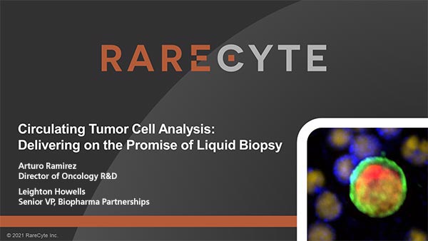
Patient Treatment Selection & Disease Monitoring by CTC enumeration
Speaker
Arturo Ramirez
Director of Oncology R&D
Leighton Howells
Sr VP, Biopharma Partnerships
Recent developments in tissue imaging technologies have made available new tools that promise to advance our understanding of human disease and help guide the development and implementation of new therapies. This interactive webinar will focus on the use of multiplexed antibody-based tissue imaging of multiple antigens at single cell resolution using both cyclic immunofluorescence (CyCIF) and Orion high-plex imaging and the use of this data to identify cell lineages and states (molecular phenotypes) and map their spatial organization and interactions at different scales.

Resolving the Complexity of Tissue Immune Response with Orion High-Dimensional Imaging
Speaker
Eric Kaldjian, MD
Chief Medical Officer, RareCyte
High-dimensional imaging allows identification of immune cell sub-types for investigation of cell number, density, proximity, and activation state. The breakthrough Orion platform generates same-day whole-slide images with sub-cellular imaging resolution in a single stain, single scan workflow with customizable staining panels. This webinar describes the application of 17-plex immuno-oncology fluorescence panel and H&E images on the same tissue sections for investigation of the complexity of immune response in normal and malignant tissues.
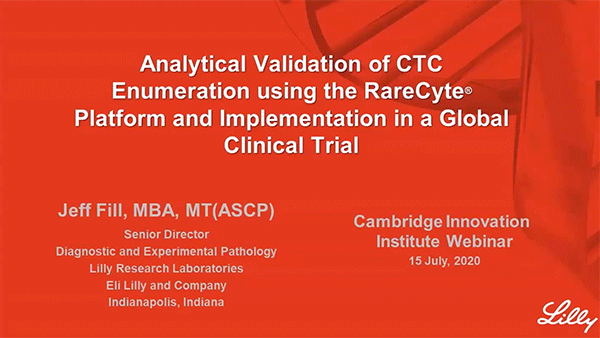
Analytical Validation of CTC Enumeration using the RareCyte Platform and Implementation in a Global Clinical Trial
Speaker
Jeff Fill, MBA, MT(ASCP)
Sr Director, Diagnostic and Experimental Pathology, Eli Lilly
This webinar describes how the RareCyte platform for CTC analysis was validated in the Clinical Diagnostics Lab (CAP accredited CLIA Lab) at Eli Lilly. The results met the requirements of the Clinical Drug Study Team and the RareCyte platform was added to a global clinical trial for further evaluation in collaboration with a CRO. A short presentation on RareCyte CTC assays for enumeration, multi-biomarker analysis, and custom assay development capabilities follows.