Webinars
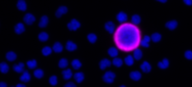
New discoveries in HER2 detection: A novel method of multimodal detection of HER2 from a single tube of blood
SpeakerEszter Papp, PhD
R&D Product Manager, CellCarta
In this webinar, Dr. Papp presents a novel method for oncosome detection developed by the CellCarta team using the RareCyte CTC platform. She also presents findings from a proof-of-concept study that combined data from oncosomes, CTCs, and cfDNA analysis to identify metastatic breast cancer patients with elevated HER2 expression.
![]()
![]()
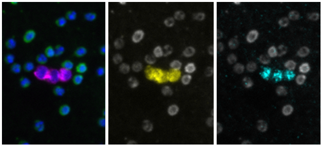
Use of open-source software for quantitative analysis of multiplex images
Speaker
Dr. Sara McArdle
La Jolla Institute of Immunology
Dr. McArdle discusses her use of open-source software for quantitative spatial analysis of tissue images. She reviews how multiplex immunostaining is a key method for understanding spatial context of the tissue microenvironment and statistical quantification of cells.
Key highlights of this webinar are how multiplex immunostaining provides:- High dimensional analysis of single samples for complex phenotyping
- Spatial relationships between targets of interest essential for studying cell recruitment, tissue development, and signaling mechanisms
- Ability to see heterogeneity within a tissue or between locations within the same tissue; important for providing spatial context
![]()
![]()
Multimodal tissue imaging and machine learning to advance precision medicine
Speaker
Dr. Peter K Sorger
Harvard Medical School
The effective treatment of cancer and many other diseases is increasingly dependent on a precision approach in which the quantification of molecular features at the level of individual patients is used to guide treatment plans. However, cancer diagnosis and staging are currently performed primarily via direct examination of biopsy and resection specimens by histopathologists. These classical methods provide insufficient molecular insight to guide the use of targeted and immunotherapies even when supplemented by knowledge of tumor genotypes.
![]()
![]()
Spatial analysis and high-plex immunofluorescence to study human pancreata in type 1 diabetes
Speaker
Dr. Estefania Quesada-Masachs
Instructor, La Jolla Institute for Immunology
In this webinar, Dr. Quesada-Masachs will explain the image analysis pipeline that she has developed and applied to study whole tissue pancreatic samples of donors with type 1 diabetes (T1D). Those samples were previously stained and acquired with the high-plex Orion™ platform. New methods for spatial image analysis are providing a better understanding of local processes occurring in many human diseases, revolutionizing those scientific fields.
![]()
![]()
Spatial Biology: Applying Cellular Phenotyping in Health and Disease
Speaker
Mickael Meyrand, Field Application Scientist, RareCyte, Inc
Tissue consists of heterogeneous cell types, each with diverse functions and functional states, where spatial organization can impact patient health status. Understanding such spatial context at the subcellular level is often challenging due to low resolution imaging, throughput, and simultaneous target assessment. Orion™ technology can resolve these challenges, providing a fast path to whole slide, multiplexed, and high resolution biomarker imaging data. Read the webinar transcription ➝
![]()
![]()
Circulating Tumor Cells Circa 2022
Speaker
Daniel E. Sabath, MD, PhD
University of Washington Department of Laboratory Medicine and Pathology
Detecting small numbers of tumor cells is technically challenging, especially for patients with solid tumors, where very small numbers of tumor cells are present in the peripheral blood, even in patients with widely metastatic disease. In this presentation, Dr. Sabath will describe various technologies for detecting circulating tumor cells and data suggesting clinical utility. In particular, his laboratory's clinical validation of a circulating tumor cell assay using density gradient centrifugation to enrich for circulating tumor cells. Dr. Sabath will also discuss possible future applications of this technology.
![]()
![]()
Webinars On-Demand
Unlock Spatial Biology with Orion Technology: Explore the possibilities for research and clinical applications
Tissue consists of heterogenous cell types, each with diverse functions and functional states, arranged spatially in a way that impacts patient health status. Resolving this complexity at the subcellular level has historically been challenged by image resolution, the number of targets that can be simultaneously assessed, and throughput. Orion™ technology breaks these barriers by providing the fastest path to whole slide, high-plex imaging which will be shown in this webinar. Read the webinar transcription ➝
![]()
![]()
Multiplexed Tissue Imaging using the Orion Platform to Reveal the Spatial Biology of Cancer
Recent developments in tissue imaging technologies have made available new tools that promise to advance our understanding of human disease and help guide the development and implementation of new therapies. This interactive webinar will focus on the use of multiplexed antibody-based tissue imaging of multiple antigens at single cell resolution using both cyclic immunofluorescence (CyCIF) and Orion high-plex imaging and the use of this data to identify cell lineages and states (molecular phenotypes) and map their spatial organization and interactions at different scales. Read the webinar transcription ➝
![]()
![]()
Patient Treatment Selection & Disease Monitoring by CTC enumeration

Join our expert panelists and learn from their experience utilizing the RareCyte platform for CTC enumeration, biomarker expression and single cell sequencing. Specifically addressing how CTC detection is possible in multiple types of GI tumors such as colorectal, esophageal, cholangiocarcinoma and pancreatic cancer.
![]()
![]()
Circulating Tumor Cell Analysis: Delivering on the Promise of Liquid Biopsy
CTC analysis via liquid biopsy (blood) allows for a less-invasive method to identify cancer cells and can prove helpful in prognosis, treatment selection, patient stratification, and patient monitoring. Our two distinguished speakers, with over 50 years of combined experience in the field, will walk through these applications and supporting data.
![]()
![]()
Resolving the Complexity of Tissue Immune Response with Orion High-Dimensional Imaging

High-dimensional imaging allows identification of immune cell sub-types for investigation of cell number, density, proximity, and activation state. The breakthrough Orion platform generates same-day whole-slide images with sub-cellular imaging resolution in a single stain, single scan workflow with customizable staining panels. This webinar describes the application of 17-plex immuno-oncology fluorescence panel and H & E images on the same tissue sections for investigation of the complexity of immune response in normal and malignant tissues.
![]()
![]()
Analytical Validation of CTC Enumeration using the RareCyte Platform and Implementation in a Global Clinical Trial
This webinar describes how the RareCyte platform for CTC analysis was validated in the Clinical Diagnostics Lab (CAP accredited CLIA Lab) at Eli Lilly. The results met the requirements of the Clinical Drug Study Team and the RareCyte platform was added to a global clinical trial for further evaluation in collaboration with a CRO. A short presentation on RareCyte CTC assays for enumeration, multi-biomarker analysis, and custom assay development capabilities follows.
![]()
![]()