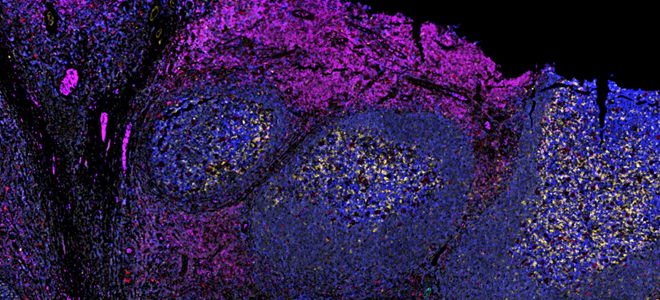
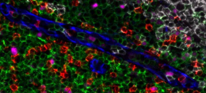
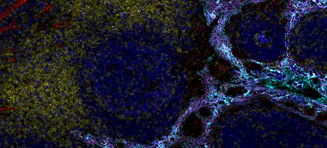
Precision Spatial Biology with Orion Multiplex Immunofluorescence Imaging
Orion is a benchtop, high resolution, whole slide multimodal imaging instrument. A combination of quantitative immunofluorescence and bright field imaging enables robust spatial biology studies and biomarker quantitation.
Overview of Orion Multiplex Immunofluorescence Imaging
Utilizing a swift sample-to-data workflow, all channels are acquired across the entire slide in one scan while rapid data processing enables whole slide, unbiased quantitation. Quick and precise sample-to-data speed provides more time for analysis and less time spent setting up your experiment. Save even more time by using our already validated panels with the standard IHC tools you already have on your workbench.
Orion: Precision Spatial Biology. Download the product brochure.
- Single cycle, whole slide staining and scanning maintains tissue integrity
- Fast and flexible panel design for rapid, robust assay validation and execution
- Quantitative data across a broad dynamic range and subcellular resolution
Case study: Understanding Disease with Multiplex Imaging and Spatial Analysis
Learn how Orion was used to understand local processes occurring in human disease in this webinar presented by Dr. Estefania Quesada-Masachs of the La Jolla Institute for Immunology.
Spatial analysis and high-plex immunofluorescence to study human pancreata in type 1 diabetes
Dr. Estefania Quesada-Masachs
Instructor, La Jolla Institute for Immunology, La Jolla, CA
Watch the webinar ➝
![]()
![]() See Orion citations on Google Scholar
See Orion citations on Google Scholar
Orion delivers fast and reliable precision spatial biology
- High fidelity data enables actionable insights
- One step, whole slide staining and scanning
- Compatible with clinical and archived samples
- Subcellular resolution and biomarker quantitation across a broad dynamic range
- Analyze TMAs or whole sections of any size
- Throughput for translational and clinical studies

Orion instrument specifications
- Full-slide scan in 75 minutes per cm2 at 20x
- Flexible panel design – use your own antibodies
- Same slide immunofluorescent and H&E imaging
- Use standard slides and sample preparation
- Easily fits on a standard laboratory benchtop
For comprehensive specifications, download the Orion specification sheet.
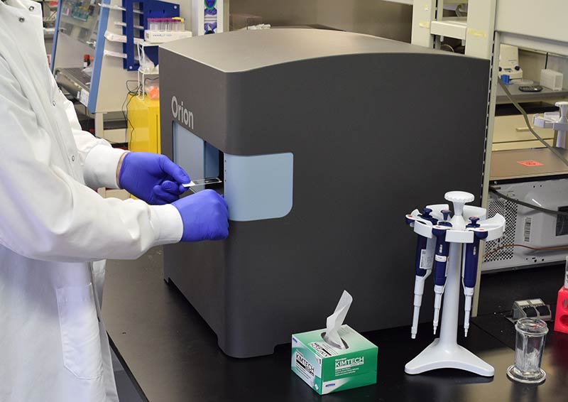
Orion's compact design makes it ideal for any lab benchtop
Orion software
Orion’s on-board software provides database functionality for easy file management and enables powerful image visualization, annotation, and sharing to facilitate collaboration for highly multiplexed datasets. Users also have the flexibility to export quantitative data to the pipeline of their choice.