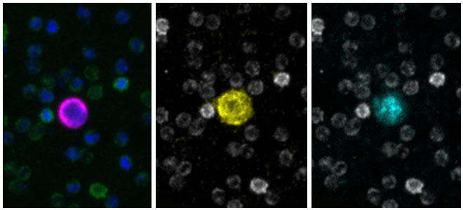
Immunofluorescence Imaging for Rare Cell Detection
CyteFinder® II Instruments are high-speed, whole slide imaging systems with options for multiplexed liquid biopsy and tissue spatial analysis.
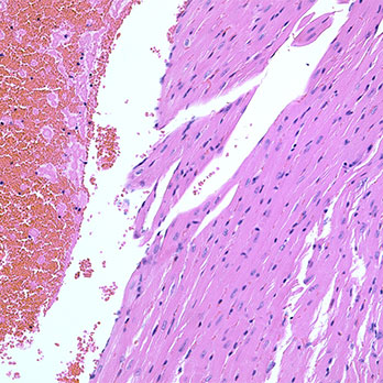
- IF, H&E, and IHC modes for imaging blood smears, tissue sections, and cytology preparation including fine needle aspirates
- Integrated machine learning algorithm detects and rank orders candidate cells for user review
- Tissue option for digital pathology on the CyteFinder II Instrument
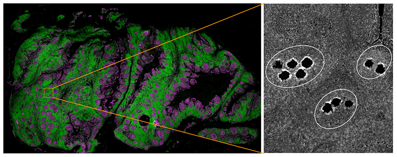
- CytePicker® Retrieval Module enables single cell or tissue micro-region retrieval from any location on the slide (CyteFinder II Instrument only)
- Designed for placement in clinical laboratories and compatible with custom analysis workflows
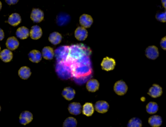
Applications include:
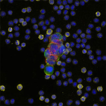
- CTC-based liquid biopsy
- Rare cell identification
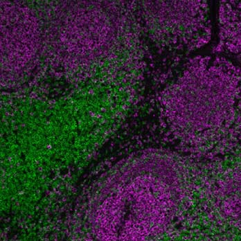
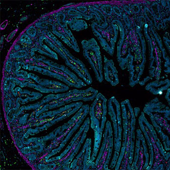
- Multiplexed, high resolution tissue imaging
- Discovery biology including investigation of tissue micro-environments
- T-cell antigen receptor discovery
- Immune cell characterization
- High volume slide imaging for digital pathology and liquid biopsy analysis
- Cell-based non-invasive prenatal testing
Case Study
Accurate CTC detection is critical in the recovery of useful, quantitative data from patient blood. A research article published in Frontiers in Oncology describes the AccuCyte process for CTC isolation and CyteFinder II’s immunofluorescence staining and imaging, together providing a reliable and accurate workflow at > 95% when compared to recovery rates using density or lysis-based processes.

Accurate isolation and detection of circulating tumor cells using enrichment-free multiparametric high resolution imaging
Yeo D, Kao S, Gupta R, et al.
University of Sydney, Sydney, Australia
![]() See CyteFinder citations on Google Scholar
See CyteFinder citations on Google Scholar
Additional articles and research
Explore CyteFinder II imaging in a high-definition map of melanoma as part of the Human Tumor Atlas Network – a National Cancer Institute funded Cancer Moonshot initiative.
The Spatial Landscape of Progression and Immunoediting in Primary Melanoma at Single-Cell Resolution
Nirmal A, Maliga Z, Vallius T, et al.
Harvard Medical School, Boston, MA
Dr. David Rimm from Yale School of Medicine discusses the importance of the next-generation CLIA diagnostic tests and HER2 positive breast cancer patients.
Watch the Yale ground rounds video ➝
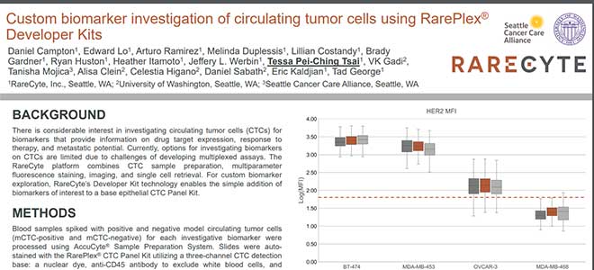
Quantitative measurement of HER2 expression to subclassify ERBB2 unamplified breast cancer
Moutafi M, Robbins C, Yaghoob V, et al.
Yale School of Medicine, New Haven, CT
Explore CyteFinder II imaging in a 3D atlas of colorectal cancer, created by the teams at Brigham and Women’s Hospital, Harvard Medical School, and in collaboration with investigators at Vanderbilt University.
Multiplexed 3D atlas of state transitions and immune interaction in colorectal cancer
Lin J, Wang S, Coy S, et al.
Harvard Medical School, Boston, MA
Vanderbilt University School of Medicine, Nashville, TN
Brigham and Women’s Hospital, Boston, MA
RareCyte Tools for Automation and Retrieval
Automation
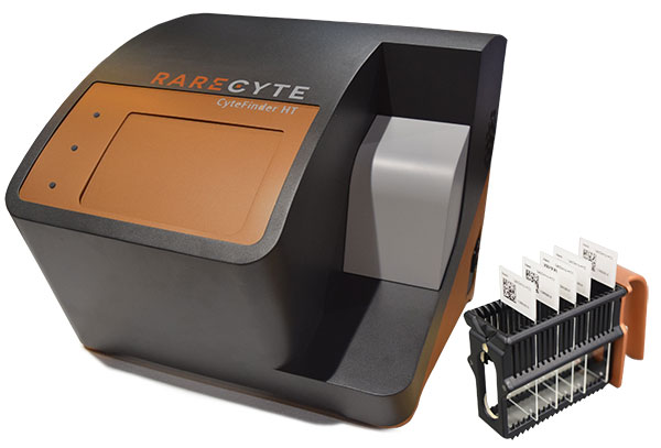
CyteFinder II HT Instrument
Scan up to 80 slides unattended
Retrieval
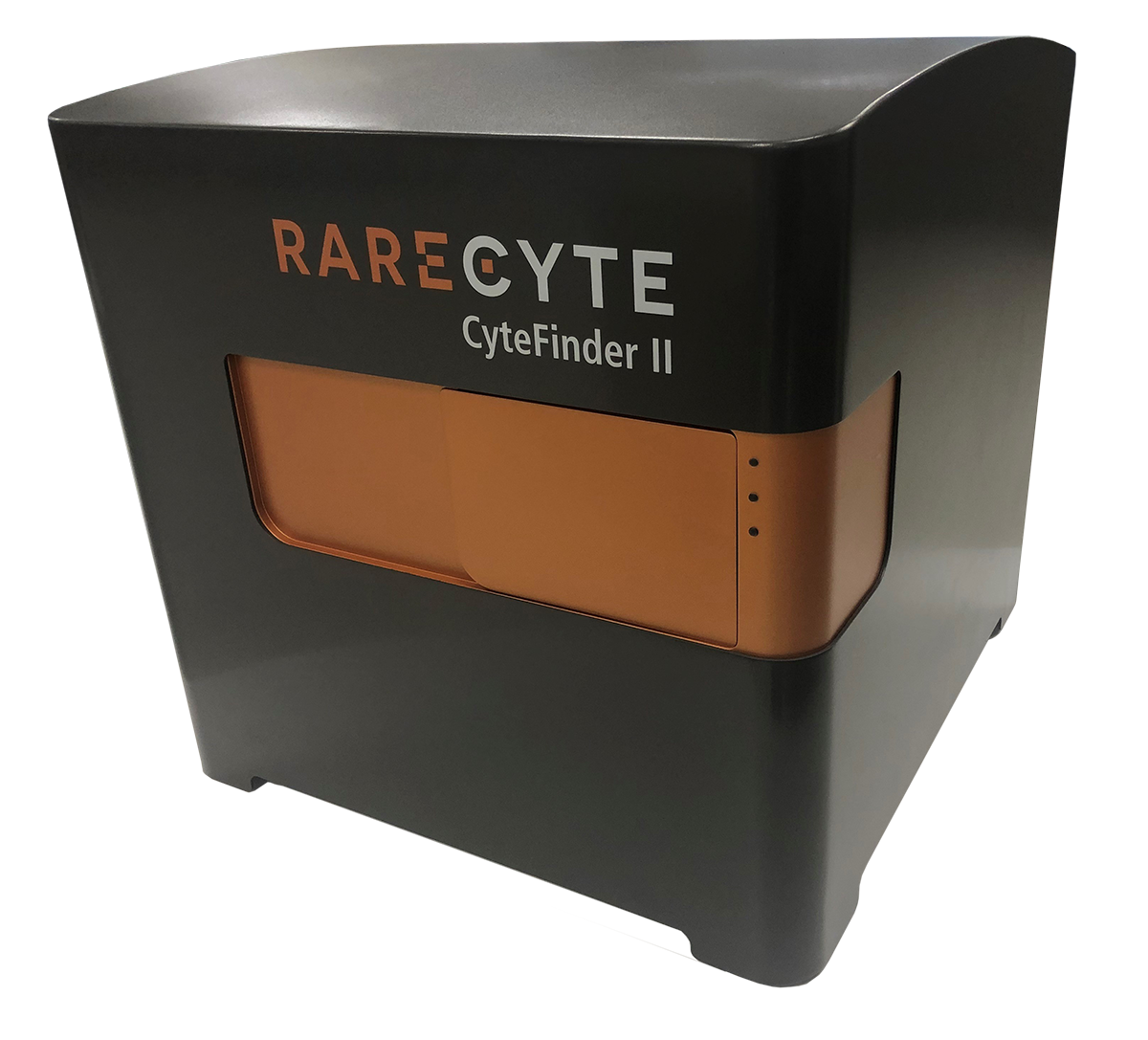
CyteFinder II Instrument
Retrieve single cells or tissue micro-regions with the CytePicker Retrieval Module
RareCyte: Your end-to-end solution for Liquid Biopsy and Immunofluorescence Imaging and Analysis
RareCyte is a full-service provider of Precision Biology™ services utilizing best in class platforms for both spatial biology and liquid biopsy. We deliver quality-focused, end-to-end services from custom assay development to clinical trial testing for researchers, translational scientists, and drug development and diagnostics organizations.
Our CLIA- certified laboratory delivers full-service programs to support all aspects of rare cell analysis from the development of protein based custom multi-biomarker assays to single cell molecular analysis. Our end-to-end platform from sample collection to result delivers exquisitely sensitive, specific, accurate, and reproducible data.
To order product, please visit our product order page.


 I would like more information
