Orion™
Panels & Biomarkers
Simple, Flexible Spatial Biology Panel Design
Easily mix and match biomarkers for your panel
Orion panels are flexible and easy to develop. Build one from a list of 150+ validated biomarkers or add your own custom biomarkers. If you aren't sure where to start, use one of our verified panels.
The Orion platform offers flexible, customizable off-the-shelf panels and supports custom panel development, making it a powerful solution for high-plex spatial biology research with over 150 validated biomarkers and dedicated support for custom antibody generation using ArgoFluor™ conjugation kits
Verified and Customizable Panels
Orion offers 16 human and 6 mouse ready-to-use panels designed for core research areas such as immuno-oncology and cellular phenotyping, with each panel easily customizable to add or remove biomarkers as needed.
Download the Orion Panel Kits List ➝
More Than 150 Validated Biomarkers
Orion features a catalog of rigorously validated biomarkers for both human and mouse applications, ensuring robust, reproducible results across diverse tissue types.
- All Orion antibody-conjugates are extensively IHC-validated and optimized for high-plex spatial imaging on the Orion platform
- Verified panels span key biological pathways, cell types, disease models, and research applications
Download the Orion Biomarkers List ➝
ArgoFluor Conjugation Kits for Antibody Customization
- Kits include dyes and reagents for simple conjugation and labeling of primary antibodies
- Conjugated antibodies can be easily validated for performance on Orion, ensuring consistency and reproducibility
NEW
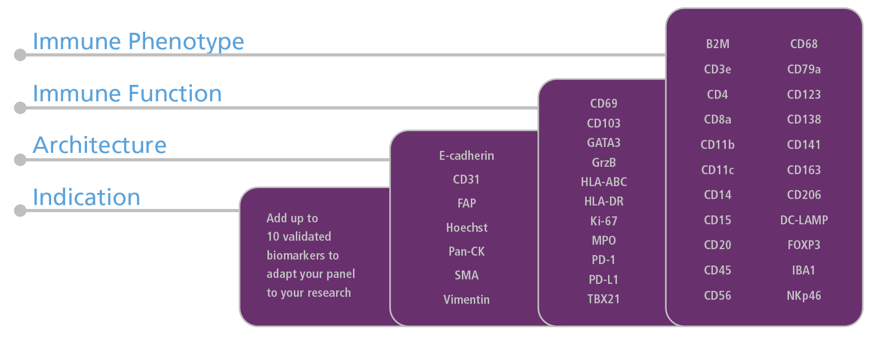
Immune Profiling Panel
The Immune Profiling Panel gives research teams both depth and flexibility. With 40 core biomarkers spanning immune phenotype, function, and tissue architecture, you can generate spatial insights immediately. Tune the panel by expanding it up to 50 biomarkers in 3 imaging rounds to adapt to your research needs.
Orion Panels for Spatial Applications
Orion panels span multiple unique tissue types, including whole tissue sections and tissue microarrays, and are utilized across multiple applications, such as Oncology and Autoimmunity indications.
