Interactive Multiplex Tissue Imaging Data Sets
These whole slide multiplex data sets show the transformational power of the Orion™ system, enabling comprehensive phenotypic profiling and characterization of tissue architecture, tumor heterogeneity, and the immune response. Contact us to learn more →
High-Plex IF + H&E Imaging of a 40-Patient Colorectal Cancer Cohort
This data set describes an approach for collecting and analyzing H&E and high-plex immunofluorescence (IF) images from the same cells in a whole-slide format suitable for translational and clinical research and eventual deployment in diagnosis. Highly multiplexed tissue imaging, spatial statistics, and machine learning were used to identify cell types and states underlying morphological features of known diagnostic and prognostic significance in colorectal cancer.
15-plex Orion™ Imaging of Breast Adenocarcinoma
This is a whole-slide tissue with same section H&E imaging of a high-grade breast ductal adenocarcinoma. The tumor is infiltrated by T cells and macrophages.
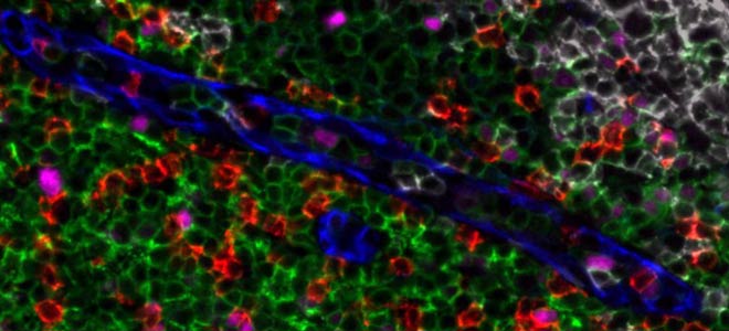
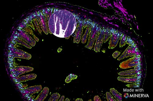
15-Plex Orion™ Imaging of Mouse Ileum
This is a whole-slide tissue section of a mouse ileum stained with a 15-plex biomarker panel and imaged with the Orion instrument in a single staining and scanning process.
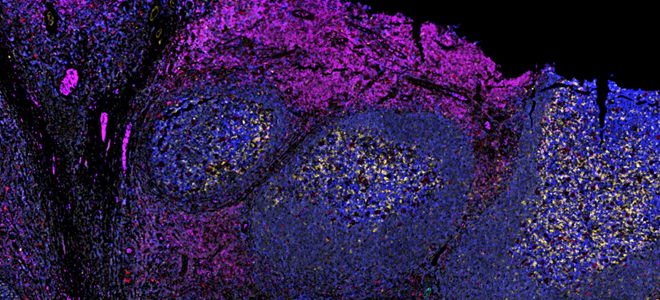
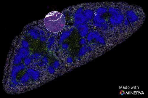
13-Plex Orion™ Imaging of Mouse Spleen
This is a whole-slide tissue section of a mouse spleen stained with a 13-plex biomarker panel and imaged with the Orion instrument in a single staining and scanning process.
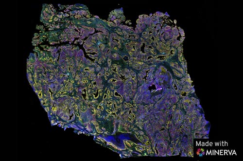
14-Plex Orion™ Imaging of Liver with metastasized CRC
This is a whole-slide tissue section of a liver sample stained with a 14-plex biomarker panel and imaged with the Orion instrument in a single staining and scanning process. Image courtesy of Ankur Sharma, Harry Perkins Institute of Medical Research
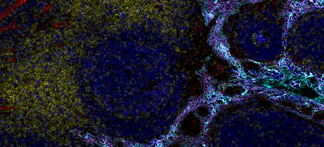
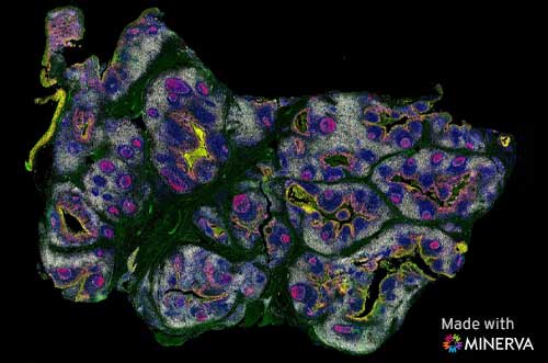
15-Plex Orion™ Imaging of Tonsil Lymphoid Hyperplasia
This is a whole-slide tissue section of a tonsil with reactive lymphoid hyperplasia stained with a 15-plex biomarker panel and imaged with the Orion instrument in a single staining and scanning process. Images in this data set show subsets of markers that have been selected from the larger immuno-oncology panel.
17-Plex Orion™ Imaging of Colorectal Adenocarcinoma
This is a whole-slide tissue section of an invasive colorectal adenocarcinoma stained with a 17-plex Immuno-oncology biomarker panel and imaged with the Orion system in a single staining and scanning process. This data was collected in collaboration with the Harvard Laboratory of Systems Pharmacology. Images in this viewer show smaller sets of markers that have been selected from the larger panel.
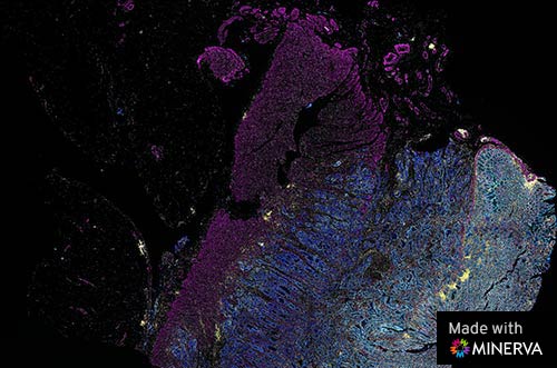
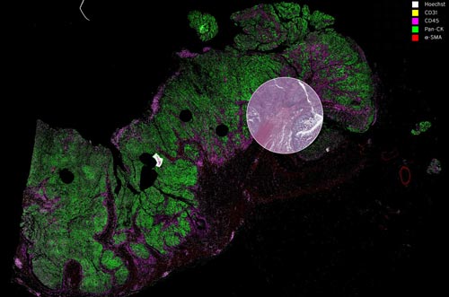
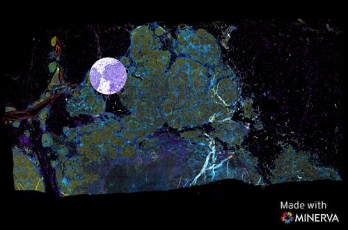
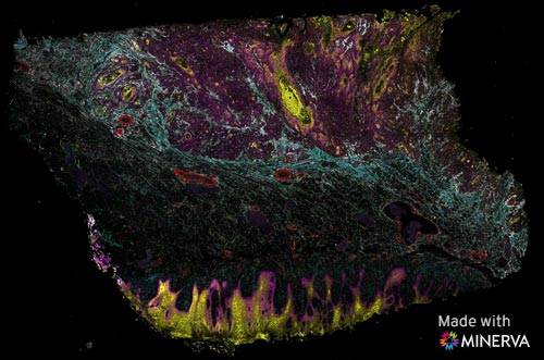
16-Plex Orion™ Imaging of Oral Squamous Cell Carcinoma
This data set displays 16-markers on a single tissue section. The normal mucosal surface of the tongue is at the bottom of the image and the nodule of invasive cancer is seen in the top half, surrounded by fibrous reaction. The slide was stained with Orion reagents and imaged using the Orion Instrument. All markers were stained in a single staining procedure and imaged in a single scan.
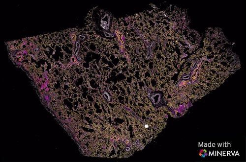
17-Plex Orion™ Imaging of NSCLC-adjacent Lung
This data set is a 17-marker whole-slide scan of a single section of lung from a region adjacent to a non-small cell lung carcinoma. It has intact normal lung alveolar architecture, with scattered immune cell infiltrates. The slide was stained with Orion reagents and imaged using the Orion Instrument. All markers were stained in a single staining procedure and imaged in a single scan.
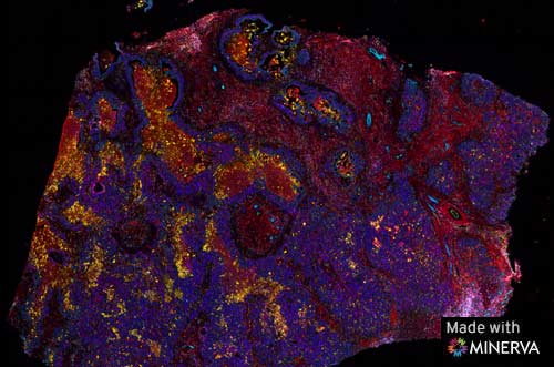
13-Plex Orion™ Imaging of Colon Adenocarcinoma
This data set is a 13-marker whole-slide tissue imaging of a high-grade colon adenocarcinoma. This is a hot tumor that contains prominent clusters of PD-L1 expression scattered throughout. All markers were stained in a single staining procedure and imaged in a single scan.
How Can Multiplex Tissue Imaging Improve Research?
Multiplex tissue imaging has emerged as a transformative tool in biomedical research, offering significant advantages for investigating complex biological processes and disease mechanisms. This advanced imaging technique enables the simultaneous visualization of multiple molecular markers within a single tissue sample, providing researchers with a comprehensive and spatially resolved view of cellular interactions and protein expression patterns. Such capabilities have profound implications for improving research outcomes.
Highly multiplexed biomarker quantitation provides a detailed understanding of cellular heterogeneity within tissues, aiding in characterization of cell types and their functional states. This is valuable in cancer research where the tumor microenvironment is highly dynamic. Multiplex imaging facilitates identification of novel biomarkers and potential therapeutic targets by revealing intricate molecular networks.
High resolution, full slide imaging enhances our ability to study a range of therapeutic areas including immunology, immune-oncology, neuroscience, and infectious diseases. By providing a holistic view of tissue architecture and molecular events, it contributes to a deeper comprehension of disease pathogenesis and the development of more targeted and effective therapeutic strategies.
Orion multiplexed immunofluorescence imaging and quantitation represents a pivotal advancement in research methodologies, empowering investigators to glean richer insights into complex biological phenomena and ultimately improve human health.