Developer Kits – 405/488
Download Specification Sheets
Download Test Menu Catalog
RarePlex® Synaptophysin CTC Marker Panel Kit
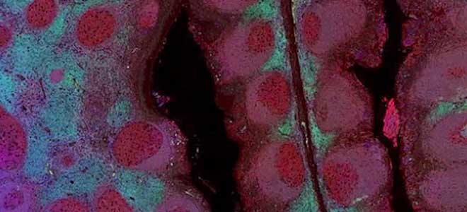
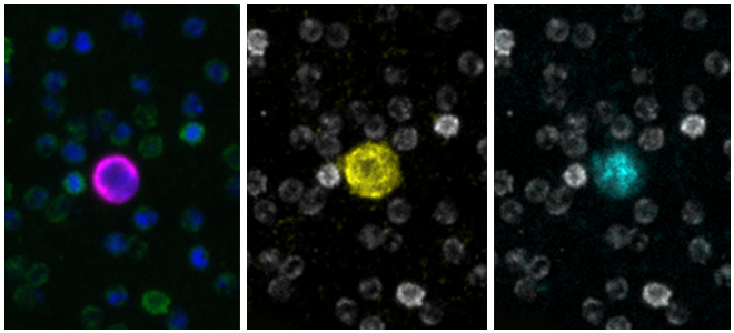
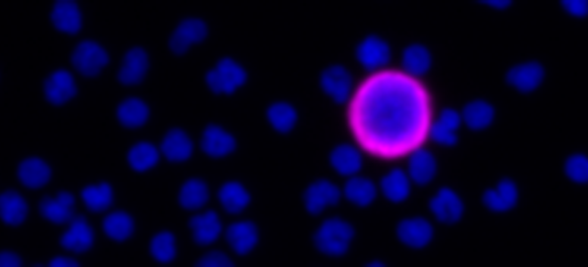
Tumor expression of Synaptophysin in cancer is an indicator of neuro-endocrine differentiation, a prominent mechanism by which tumors become resistant to therapies. The RarePlex 0920-VB Synaptophysin CTC Panel Kit enables immunofluorescent detection of Synaptophysin marker (SYP) expression by epithelial circulating tumor cells (CTCs). Analysis of CTCs via blood allows for a less-invasive method to identify cancer cells and can prove helpful in prognosis, treatment selection, patient stratification, and monitoring.
With the flexibility of one open channel, the RarePlex Synaptophysin CTC Panel Kit expands clinical research of therapeutic resistance by combining SYP with any other marker of your interest. This assay is designed for use with the AccuCyte® Sample Preparation System and the CyteFinder® Instrument for a blood-to-result solution.
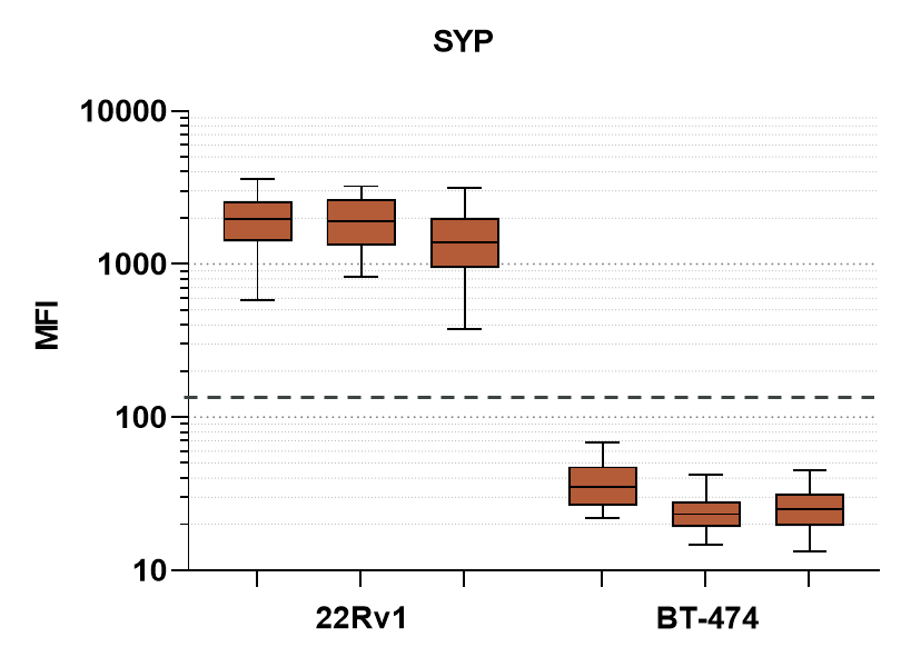
Analytic validation studies* using spike-in SYP+ and SYP- cancer cell lines as model CTCs (mCTCs) have demonstrated the RarePlex Synaptophysin CTC Panel Kit to be highly specific, sensitive and accurate:
- SYP biomarker Accuracy of 99%
- SYP biomarker Sensitivity of 99%
- SYP biomarker Specificity of 100%
*Studies were performed using quantitative mean fluorescence intensity measurements of mCTCs
What is included in the Synaptophysin CTC Panel Kit
The kit contains a nuclear dye; a panel of antibodies and reagents for detection of CD45 (to exclude WBC), Cytokeratin and EpCAM, and Synaptophysin; instructions and protocols for automated staining and scanning; and guidelines for interpreting results.
Why is Synaptophysin Important?
Synaptophysin holds significant importance in the realm of circulating tumor cell (CTC) research. By detecting synaptophysin expression in CTCs, researchers gain insights into their neuroendocrine origin and potential metastatic behavior. As synaptophysin is a biomarker associated with neuroendocrine tumors, its presence in CTCs can aid in diagnosing and monitoring these malignancies. Furthermore, studying synaptophysin expression in CTCs can provide valuable information about tumor aggressiveness and treatment response, assisting in personalized treatment decisions. The assessment of synaptophysin in CTCs offers a non-invasive approach to evaluate tumor characteristics and dynamics, facilitating the development of targeted therapies and improving patient outcomes in the context of CTC research.
How biomarkers are used in clinical research?
Biomarkers, like synaptophysin, are integral to clinical research, enabling early disease detection, prognosis prediction, and treatment monitoring. They provide insights into disease mechanisms, aid in the development of targeted therapies, and facilitate personalized medicine, enhancing patient care and advancing medical knowledge.
Additional resources for the RarePlex Synaptophysin Panel Kit
Download the specification sheet →
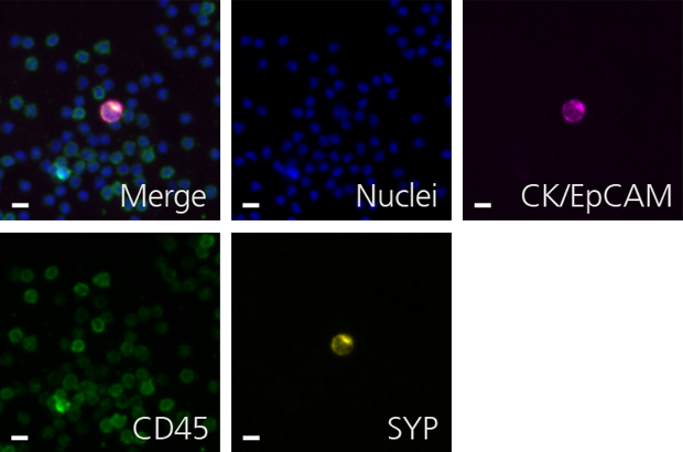
Scale bar represents 10μm.